
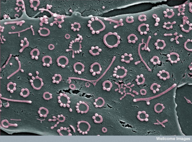
SMALLEST CELL: Mycoplasma. The pink circles are a type of bacterium only 0.1 μm in diameter (0.0001 mm, or about 0.0000039 inches).
• They have the fewest known genes of any free living organism. Mycobacterium laboratorium is synthetic bacterium patented by Craig Venter, who has whittled down the minimum number of genes to 382.
• Lacking a cell wall, they are immune to common antibiotics like penicillin that target cell wall synthesis. M. pneumoniae and M. genitalium cause human disease.
• In the lab, they are a common contaminant of cell culture and can seriously confound results. Difficult to detect unless one specifically looks for them, mycoplasma contamination is estimated at minimum of 10%.
Scanning Electron Micrograph: Kevin MacKenzie, University of Aberdeen, Wellcome Images. Mycoplasma on surface of bone-forming osteoblast cells.
#scienceeveryday

My friend you live in a fascinating world …:)
Also, they can cause real bad contamination in tissue culture…
Wonderful image 🙂
Scary, too Kershaw Rustomji . My student told me yesterday that her cell cultures were contaminated with this bug. It can really mess up experiments.
Ah!
That is true Rajini Rao …it is almost like going into Space…where everything is unknown
Heh. Software enthusiasts whittle their code down to bytes and show it off in the demoscene. Biologists get a patent.
Craig Venter is somewhat unusual, Richard Healy ! Perhaps the Steve Jobs of the computing world?
Yes indeed Rajini Rao !
It was an amazing feat!
That’s a nice micrograph. Pretty colours. I’m looking forward to the first cell being completely synthesised from scratch. Mycobacterium laboratorium was kind of a cheat in that they used the cytoplasm of an existing cell and only synthesised the DNA. Still a major accomplishment, of course. But synthesising the DNA, the cytoplasm, and the cell wall from raw elements will finally show that life is not some kind of Godly creation, it’s merely very complicated biochemistry.
nice
I’m not a Venter fan, Drew Sowersby . He gave a terrible talk when he visited Hopkins right after the Human Genome publication..it sounded like a spiel to venture capitalists rather than to his peer scientists, many who actually contributed to the human genome (separately). Although I did laugh at his parking his car (I think it was a red Ferrari or something flashy) illegally in east Baltimore (not the safest neighborhood).
Synthesizing DNA is relatively easy. I don’t see a way to synthesize cytoplasm or the cell wall! Based on existing technology? Nope!!
Recreating the _exact_ milieu of the cytoplasm seems impossible. For that we would have to know the exact amount of all the cellular enzymes, sugars, organelles, and what not!
Is that the reason Rajini Rao ?
WhatDo EngineersDo , they sure are. I’ve never seen circles like this, I wonder why they look like that in this scan.
Are they the colonies, or individual organisms?
Deeksha Tare , exactly. Forgetting the amounts/proportions for a moment, how on earth could we synthesize ribosomes, complex membranes and their embedded proteins, and all the macromolecules that go with it. We can direct their synthesis with genes and building blocks but we still need a pre-existing cell to do it in.
Not sure, Deeksha Tare . I’m guessing they are colonies..some rounded cells and some long. But why are they arranged in circles?
Yaa! Sometimes the wondrous world of a small, tiny cell can leave you speechless!!
The kind of multitasking that goes on inside it is anybody’s guess!
They are small, Mark Bruce , just one-tenth the size of E. coli. But they pay a price in independence, they have to scavenge or parasitize a host to get a lot of the material they need.
Deeksha Tare , apparently their cell shapes are pleiomorphic. i.e., they can form tubes with bulbous extensions, rounded ends and so on. That explains the strange morphology 🙂
Is this a picture of a cell culture?
Yes, the grey cell (osteoblast) in the background was grown in culture.
Yes! many bacteria, and some viruses too are pleiomorphic.
Yeah, Mark Bruce , I do appreciate a biologist with an artist’s eye 🙂
Yaa! Using the right color combination is really essential!
While highlighting the different domains of Dengue virus E protein for one of my assignments I used the combination of pink and purple! Looked kinda girly, but the chains stood out well! 🙂
Deadly Dengue and Girly, Deeksha Tare ! Post it on G+ if you have a chance:)
Sure, if I’m able to find it!
We all were so very fed up of all those bioinformatics assignments in the previous sem that once it was all over, I just shoved my file somewhere in the dark recesses of my shelf! Hehe!! 😉
Hi Feisal Kamil
Bye Feisal Kamil 🙂
Hehe! 🙂
Incredible. Thank you, Rajini Rao for your expert commentary.
Good point, Feisal Kamil , because they should all be round in that case. Why are they elongated, I wonder? Perhaps they are adhering to the substrate host cell and growing out.
Bravo, Feisal Kamil . That’s it! Since they divide by binary fission: “The transformation of mycoplasma filaments into chains of cocci .. revealed the appearance of constrictions in the cell membrane at about equal distances along the entire length of the filament, a process that takes a few minutes. At the time these observations were discussed, one had no idea about the mechanism of this process. Recent genetic data throw some light on the genes and proteins involved in maintaining the peculiar cell shapes and the cell division process in mycoplasmas.” Also, I found that mycoplasma have a rudimentary cytoskeleton that helps maintain their shape. Thanks!!
AFAIK they are taxonomically grampositive but lack the Gram-colorable layer. So what is left of their cell wall or membrane? I think there must be something, otherwise their molecules would swim away.
Ralf Muschall , they are derived from gram positives by reductive evolution. Although they have no cell wall left, they do have a plasma membrane (like animal cells). The membrane is unusual in having a lot of sterol that they extract from host cells. Apparently this makes them particularly fusogenic, so they can infect animal cells.
These things fuel nightmares.
I am guessing that if they are so common, we prolly have many of these living inside of us at any given instant? With them being immune to antibiotics, I take it that our immune system does a pretty good job at keeping these at bay or are these things killing us slowly as we speak? :p
Edit: M. pneumoniae and M. genitalium cause human disease. > missed that bit… they really are killing us. 😦
Fascinating picture. In addition to what others have mentioned, I also find it interesting that they seem to want to arrange themselves in a rectangular grid or array. I can see how an even spacing would give everybody equal access to nutrients. I wonder if they kind of adjust their positions over the surface to find some kind of equilibrium. But I suppose that would have to keep changing as they multiply.
stefan jeffers , I had not noticed how evenly spaced out the mycoplasma are, that is unusual! Bacteria do communicate via chemicals by way of “quorum sensing”. I wonder if something like that was involved or this distribution was just chance.
Good morning, Feisal! I should read up on quorum sensing, just for fun.
No blather at all..you actually solved the puzzle of the shape that Deeksha Tare and I were rambling on about 🙂
I think that is what Stefan was alluding to, re. the spacing, but that would mean that the bacteria have a way to detect gradients of chemicals and position themselves in a way that maximizes the concentration of the particular nutrient they are sensing. Certainly, any cell can detect nutrients (particularly sugar), but I don’t know if it would move (“chemotaxis”) to place itself in optimum position. Interesting idea, long answer (sorry).
You’re right..they must do chemotaxis. They are motile..apparently have a gliding movement.
They’re growing on a mammalian cell (grey background) according to the caption, Drew. I think the osteoblast must have been placed on the EM grid, not the mycoplasma itself. That’s why it’s strange.
They can move around..not sure how much on the cell surface though..may be bound to receptors on the host membrane.
The membrane potential is always negative inside, positive outside by about 65 millivolts. That would be mostly Na+ ions near the outside of the membrane surface making it positive.
Drew Sowersby , turns out there is a large literature on receptor interactions 🙂 Toll like receptors on host and lipoproteins on mycoplasma.
http://www.ncbi.nlm.nih.gov/pmc/articles/PMC2613965/
Haha, aren’t they all?!
Hi and Bye…
Would be back after the practicals! 🙂
Can gliding bacteria measure gradients? For flagellum-driven ones, AFAIK gradients and bacterium diameters are to small for that to be possible – instead of that, they measure the concentration, swim a few microns, measure again, continue swimming if it improved, otherwise tumble (by spinning the flagellum backwards) and start in a random fresh direction. What determines the direction of a crawling bacterium?
Hello! I am back! After a day full of RNA extraction! 🙂
Great discussion! 🙂
Mycobacterium (causing leprosy and TB) is different from Mycoplasma, Feisal Kamil . It’s a rod shaped bacterium with a particularly resistant cell wall. Some grow extremely slowly (20 day for one division) and are a nightmare in their own right!
What are you going to be doing with RNA preps, Deeksha Tare ?
It is a part of our routine practical course Rajini Rao ,
We’ve stored the Japanese Encephalitis RNA which we extracted today (by TriZol method) at minus 70. Tomorrow we’d run a one-step PCR and then run the PCR product on a gel to check the purity and size of the extract.
hi Ranjini Rao, thank you for educating us, I like your posts 🙂 !!!
Thank you, KSG Krishnan 🙂
Wishing you a nuclease-free PCR/gel tomorrow, Deeksha Tare !
Hehe! Thanks a lot Rajini Rao !
Hey! Let’s play around with these and see what happens……..
Yeah, J Huntemann ! We may even learn something.
HI rameez ahmed HOW R U?
LOL, Chad Haney , he may transfer his love to you 🙂
I better change my profile pic to one with my pink Oxford and pink Mycoplasma.
PLEASE DON’T BE JEALOUS Rajini Rao HE’S JUST A FRIEND.
Be sure to pick that particular pink shade of M. genitalium.
but of course
Ralf Muschall , sorry for the late response. Chemotaxis has been studied in the slime mold Dictyostelium (they move towards cAMP to form fruiting bodies), in yeast (in response to mating factor secreted by the opposite mating type cell) and in immune cells. I have a colleague who studies this, and my recollection is that a gradient of the chemical is sensed by clustering of a small second messenger (PIP2) near the region of highest concentration. If the direction of gradient is changed, then the messenger concentration changes too (the movies of fluorescently labeled PIP2 are quite dramatic). I found this paper by Peter Devereotes, if you are interested: http://www.ncbi.nlm.nih.gov/pmc/articles/PMC2951443/?tool=pubmed
Did your friend leave? I have something. I’ll just leave this here.
Right Said Fred – I’m Too Sexy (2007 Mix)
ROFL, my son just awarded me a demerit!
video call
Chad Haney , RAJEEV is back 🙂
you who friend
Rameez, RAJEEV, and Michael can discuss how sexy they are with each other.
If he’s related to RAJEEV he must have 5 sexy wives and 2 boyfriends.
I have to take my dog out . I’ll be back for sugar plum.
Keep your video ready, Chad Haney 🙂
“We may even learn something.” Too much too late? Just thinking out loud…………………………..
Too little, and never early enough. Why such a pessimist, J Huntemann ? Don’t let the mycoplasma get you down.
When we play with organisms this small, and in jest say simple, we are approaching boundaries that I wonder are we knowledgeable enough to know all the consequences of our genetic manipulations?
“• Lacking a cell wall, they are immune to common antibiotics like penicillin that target cell wall synthesis. M. pneumoniae and M. genitalium cause human disease.
• In the lab, they are a common contaminant of cell culture and can seriously confound results. Difficult to detect unless one specifically looks for them, mycoplasma contamination is estimated at minimum of 10%.”
Where is your friend Rajini Rao
J Huntemann , the word “play” reduces a serious endeavor down to frivolous past time. It took me 25 years of schooling to acquire my own lab and that was about 10 years under average (most scientists are 40 years old before they become independently funded). To be funded to do this sort of research, one must be at the top tenth percentile of one’s peers. When others were “playing” on New Year’s Eve, I was in the cold room (walk in refrigerator) working on my project. So, no..we are not playing in any sense of the word.
Second, if one hopes to understand the human body, starting with something small and simple is the logical choice. Unless, you do not wish to understand the human body and the diseases that visit us. In that case, we can all just die of microbial infections, as we did during the 1918 influenza epidemic or the black plague.
Yes, mycoplasma do cause human disease. Isn’t that all the more reason to study them? I’m not understanding why you repeated my first bullet point. Or the second, for that matter. If it is a common contaminant, does that mean it is a sinister alien that is going to take over the planet and send us all into an early demise? That’s the stuff of Hollywood summer movies. I can’t think of such a real life man-made Frankenstein released from a research lab, can you?
I appreciate your comments. But I have a very different view of my scientific calling. I do biomedical research because I want to make a difference to the human condition, because I am passionate about science, and I am good at it. Not because I want to play God or any such nonsense.
J Huntemann what was your point of copying those sentences?
“the word “play” reduces a serious endeavor down to frivolous past time.”
I apologize for my unthinking use of the word.
” if one hopes to understand the human body, starting with something small and simple is the logical choice.”
I have enough Biology to understand somewhat the complexity of of the simplest building blocks of a life form.
I have enough math to understand the complex nature of genetic alteration.
“If it is a common contaminant, does that mean it is a sinister alien that is going to take over the planet and send us all into an early demise?”
In its wild forms, no……. and I would guess no in some of its engineered forms?
I strongly feel that one cannot account for all the results from this kind of engineered forms of this organism.
You know far better than me (just enough knowledge and education to be dangerous) to know unintended results.
“Yes, mycoplasma do cause human disease. Isn’t that all the more reason to study them?”
Yes! by all means!
” I can’t think of such a real life man-made Frankenstein released from a research lab, can you?”
Unknown.
“I do biomedical research because I want to make a difference to the human condition, because I am passionate about science, and I am good at it.”
That is an understatement if ever I heard one!
You are not good, you are preeminent! there are very few that come to your level in your study area.
I consider it an honor you responded to me. Especially after that unthinking “play” comment lead off.
Cutting to the chase, I worry allot about genetic manipulation. I worry most about that organism you picture above.
I feel strongly if something “bad” happens this will be the area that will cause the “problem” that has no solution.
It was not my intent to disparage you or your work although I did start on a bad foot!
I wanted the opinion from a scientist who is the best in her field. You.
With respect from one scientist to another,
Mark
J Huntemann (Mark), thanks for your explanation.
Unexpected consequences of genetic manipulation are legitimate causes of worry. Here is what we do to minimize them. The organism is first crippled by mutating/deleting several critical genes so that it can no longer be free living. It needs to be cultured in lab. Only then do we genetically manipulate it by putting in modified or foreign genes. We also have to add full strength bleach to the cultures before discarding, and every item of waste from labs gets incinerated.
(There are genetically modified organisms that do get released in the field of agriculture (not biomed. research). That’s a separate issue, with pros and cons.)
Mycoplasma in particular are probably less deadly than certain kinds of virus. Viruses are even smaller and simpler in organization. Speaking of modifications, viruses mutate rapidly on their own, even without our assistance. Nature is a giant laboratory and any tinkering we do pales in comparison with what goes on around us. The way I look at these bugs, is that we need to stay one (small) step ahead of them. Philosophically, it’s the same with cancer cells. It seems that the more we learn, the more complex they reveal themselves to be. The chemistry driving them to survive is both simple and powerful. Let me know if this makes sense. Cheers!
Just to add, the viruses we work with are replication deficient.
Thank you very much for your response.
I just worry because I know how tenacious life is. As you well know you find life forms in places where you would never think they could exist much less thrive.
Never rule out the impossible where life is concerned I have learned.
Again thank you for your response
Blessings
Mark
Small buggs/Bac……big problems ???
Hi
Great pic! Can you tell me what length the longest filaments are? We have something in our cells but it seems to be a bit big for mycoplasma.
Michelle Parker , it could be filamentous mold or yeast. They eventually make the culture turbid. There are mycoplasma and mold detection kits that are commercially available..we have had good results using them. Good luck!