Did you know that reindeer are the only known mammal whose eyes change seasonally from golden in the summer to a wintry blue? Their eyes also become a thousand times more sensitive in winter, adapting to dim light in the near darkness to avoid predators.
Part of this is because of changes in the tapetum lucidum (TL), a mirror-like layer under the retina.
Tapetum lucidum
The tapetum lucidum (TL) reflects light so that the photoreceptor cells in the retina get another chance of capturing stray photons. Most mammals have a golden TL, as do reindeer in the summer. But in the constant darkness of winter, muscles keeping the pupils dilated become strained and pressure inside the eye builds up (just as in glaucoma). This makes the TL flatten and packs the collagen fibers inside it so they diffract blue light.
If the reindeer are herded in regions with distant urban lights, their TL is partly altered and their eyes look green! This may not be the whole story and there could be changes in the retina as well.
A picture of a little katydid (Amblycorypha oblongifolia) crossed my time line the other day.
Katydids, named after the sound of their chirp, can be distinguished from grasshoppers and crickets by their long back legs, very long antennae and rhomboid shaped body that is tented like the roof of a house.
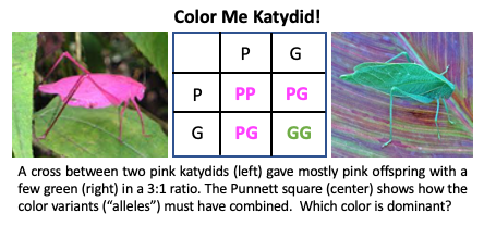
A katydid looks like a walking leaf. Nearly all of them are green, but a few are bright pink or even orange and yellow. The colors are variations of a single gene. So it appears at first sight that these rare color morphs must be recessive traits, while the common green must be the dominant variation (known as “allele”), right? Wrong!
When two pink katydids were crossed, their offspring were not all pink as would be expected if pink was a recessive allele. Instead, a few green katydids appeared, with a pink:green ratio of 3:1.
The Punnett square is used to figure out what happened in the cross. Remember that the gametes or sex cells (sperm in male and egg in female) have all their chromosomes halved, so each gamete carries only one color variant. When the sperm fertilizes the egg, the gene variants pair up in different combinations of Pink (P) or Green (G). Only the GG combination gave green katydids. Even one P was enough to result in a pink katydid.
Each of the pink katydids must have carried the recessive green trait, which was masked or silent. When mixed together, a few offspring carried two green alleles and appeared green.
But why are most of the katydids found in nature green?
This is due to natural selection. Pink katydids are conspicuous in nature and easily found by predators. More green katydids survive because they are nicely camouflaged among the leaves.
Have you ever seen a pink katydid? If you do, be sure to congratulate it on surviving the odds of natural selection. And think about how not all dominant traits are “good” for survival of the species.
And then there were two! Watch this mesmerizing time lapse video of a desmid dividing. Desmids are single celled, microscopic algae that are beautifully symmetrical. Each cell has two half-cells connected in the middle by the cell nucleus.
Did you know? Desmids thrive in clear, nutrient-poor and unpolluted fresh water. They are considered an “indicator species” of water quality because they disappear when water turns murky.
Did you know that smell receptors are sniffing out cues and signals all over our body, not just in the nose? Watch Jen Pluznick, my lab neighbor at Johns Hopkins University, explain the weird facts about smells in this TEDMED talk.
Do you have a gut feeling that this could be important? You could be right! Jen has discovered smell receptors in the kidney that appear to be responding to chemicals from gut bacteria that end up regulating blood pressure. Read more on the story here: https://goo.gl/AyBFE6
The first was on acid-base regulation and proton transport, 49th in a series that was first organized by legendary Danish scientists Hans Ussing and Nobelist Jens Skou. The Ussing chamber is a classical apparatus used to measure electrical current across a layer of cells known as epithelium, as a proxy for the ions that are transported in and out of the cells. Skou discovered one of the most important of these transporter proteins, known as the sodium pump. The meeting was held at the historic Sandbjerg estate, near Sonderburg, which dates back to the 16th century and eventually ended up with the family of author Isak Dinesan (real name, Karen Blixen) of Out of Africa fame (http://www.sandbjerg.dk/en/). It is now owned by Aarhus University, to the enjoyment of lucky researchers! The second conference was to celebrate the achievements of a colleague, Poul Nissen, structural biologist extraordinaire, of Aarhus University. Poul received the Novo Nordisk prize for his beautiful atomic structures of ion pumps, including the sodium pump that was discovered by Jens Skou. We stayed at Norsminde Kro (Kro=inn) and the science talks were at Mosegaard museum. Back to Aarhus, where the sun barely sets, before heading back home.
Yesterday, on Earth Day, tens of thousands of scientists and science enthusiasts across the world took to the streets to march for science in an unprecedented show of solidarity. We came wearing white lab coats, pink knit brain caps and costumes. We sang, chanted and cheered. We carried signs that were prophetic, political, nerdy, funny, witty and even obscure. Here are some of my favorite signs and photographs from various marches. Thanks to Chris Robinson for marching with me in Chicago, where we were both attending our respective science conferences, and taking some great photographs. Tell us if you marched (and where) and feel free to post photos of your favorite signs in the comments!
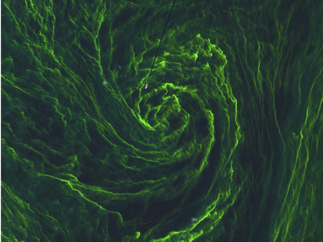
Algal Art What is the mysterious 3D whorl in this latest addition to Art or Science ? Look closer and there seems to be a scratch in top center..and is that a white speck of dust marring your monitor?
You may be surprised to learn that these delicate green swirls are an aerial view of a giant algal bloom floating in the Baltic Sea, captured by the orbiting satellite Sentinel-2A. The white speck heading into the “eye of the storm” is a ship. You can see the ship’s “wake”, caused by the propeller’s cutting through the floating algae as a straight dark line.
Annie, Fannie and Mike: They seem friendly enough, but these are actually nicknames for three types of cyanobacteria that account for the vast majority of algal blooms world-wide: Anabaena, Aphanizomenon, and Microcystis. Caused by eutrophication of water from fertilizer dumping, what could be bad about these temporary blooms of harmless sounding photosynthesizing microorganisms? “Annie” and “Fannie” produce toxins that attack your nervous system. “Mike” makes microcystin, one of the most potent toxins on the planet. Even inhaling a few droplets of contaminated water can make you nauseous and dizzy, and larger doses kill. They grow best in warm water with lots of nutrients. Thanks to warming climate and fertilizer run offs, algal blooms are on the rise, starting as early as March and April.
As algal blooms grow, others die. Bacteria divide quickly, using up the oxygen supply. Fish and aquatic life are starved of oxygen. This leads to dead zones. Scientists are combating algal blooms through innovative strategies. One way is artificial destratification by mixing up upper, warm layers with deep, cooler water using propellers. This effectively starves algae and cyanobacteria of nutrients and light. Another way is biomanipulation by introducing aquatic plants that compete with algae or predatory fish that eat other plankton eating fish. Sadly, support for this research is at an all time low. That’s why we #MarchForScience today. Show your support for #EarthDay and support science!
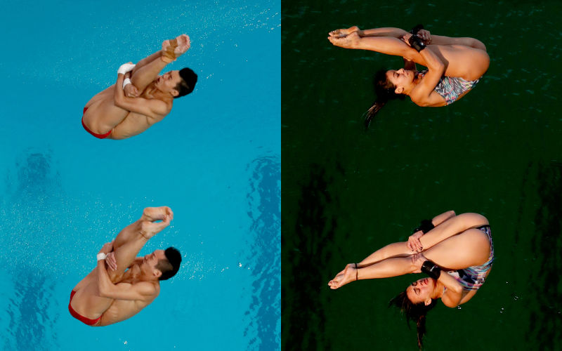
♒ We know that green is Brazil’s favorite color, and the Olympics are trying to Go Green for the environment, but even so, the overnight change in color of the Olympic swimming pool from an azure blue to murky green took scientists and sportsmen by surprise. While officials hastened to assure athletes that the green waters posed no health threat, the mystery caused much speculation. Caipirinha-flavored Soylent? Stiffed by Trump’s pool cleaning service? Who peed in the water?
♒ “Midafternoon, there was a sudden decrease in the alkalinity in the diving pool, and that’s the main reason the color changed,” said Mario Andrada, a Rio 2016 spokesman. So, the pool became more acidic. But acidic water is not green. There are two likely explanations: first, excess copper in the water can turn it green, but not murky. The latter is caused by a sudden and rapid growth of algae, triggered by the warm weather, lack of wind, insufficient chlorine and ineffective filters.
♒ Algal spores can enter the water inadvertently, carried by wind, rain and contaminated swimsuits. When the conditions are right, they can “bloom” overnight. Because these algae are visible only under the microscope, there must be millions of them in the water to change the pool color from blue to green. One way to deal with them, after normalizing the pH, is *superchlorination*—aka shocking them with high levels of chlorine. Not all the Olympians are complaining: Canadian divers said that the contrast with the sky helped them win the bronze.
♒ Pix: The Olympic diving pool on August 8 (left) and the Olympic diving pool on August 9 (right) Image: AP
⦿ Jumping spiders (Salticidae) don’t use a web to catch prey. Instead they locate, stalk and mount a jumping ambush when they are 1-2 cm away. To do this, they need to detect and then evaluate objects so they don’t confuse a potential mate as prey! Fortunately, jumping spiders have among the sharpest vision among invertebrates.
⦿ Unlike insects, spiders don’t have compound eyes. Instead their 8 “simple” eyes point forward (for high focus) and sideways (to detect motion). Strategically, this is similar to the division of labor in our eyes: we detect peripheral vision at the edges of our retina with low resolution but wide field of view, and sharp images at the fovea in the center of the retina, which is packed with a high density of vision receptors, but has a limited field of view. Since the spider’s large central eyes are set close together and have a limited field of view, they must be moved to point the fovea towards the object. How do they do this?
⦿ Involuntary leg movements are triggered by stimuli from the lateral eyes to reposition the body. However, the spider cannot swivel its whole eyeball as we do, because the lens is built into the carapace, or outer skeleton. Instead, a set of six muscles moves the retina: up and down, sideways and rotationally, while the lens stays fixed. In a transparent spider, you can see the unusual movements of the retina in the tube-like principle eyes. Just one more addition to the cuteness quotient of these tiny spiders!
Red, White and Grape: From Jumping Genes to Wrapping Leaves
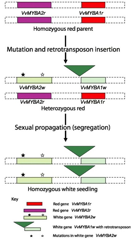
Red or White? Even King Tutankhamun (1332-1322 B.C.) prudently stashed away amphorae of both red and white wines to enjoy in the afterlife. Biochemically, a single class of pigments found in grape skin, the anthocyanins, separates the red from white. White grapes arose from their wild, dark berried ancestors by not one, but two rare and independent genetic events: either one alone would not have given us the white grape. In fact, all ~3000 white cultivars today carry these same gene disruptions, pointing back to a common ancestor that arose millennia ago. The disrupted genes code for transcription factors, aka master regulators of biochemical pathways that can turn other genes on or off.
Science sleuths have peeked back into the gene history of Vitis vinifera to figure this out.
First, the MybA gene duplicated, giving two side-by-side copies, both active in making anthocyanins and red berries. Somewhere along the way, one of them, the MybA2 gene accumulated two mutations (depicted as stars) that rendered the resulting protein non-functional.
Independently, a “jumping gene” or retrotransposon, (green triangle) landed within the adjacent backup gene MybA1, knocking it out as well. The resulting plant, termed heterozygous, still bore red berries, because the unmutated genes on the other chromosome were active. Eventually, two heterozygous plants bred together and some offspring received both chromosomes with two nonfunctional MybA genes.
Voila, white grapes!
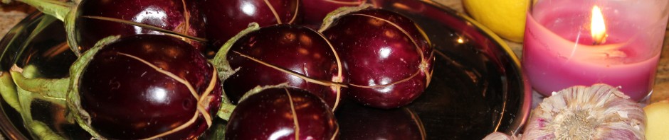
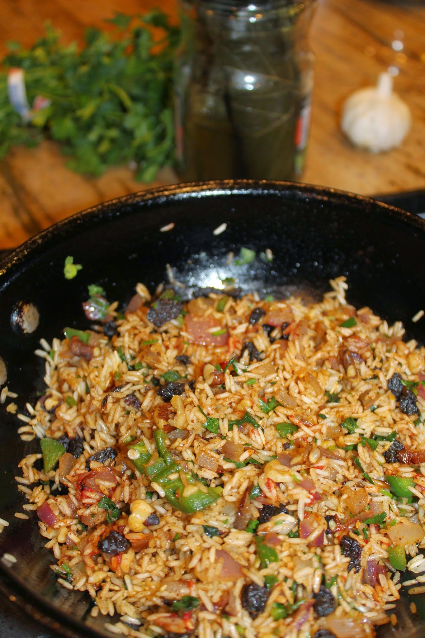
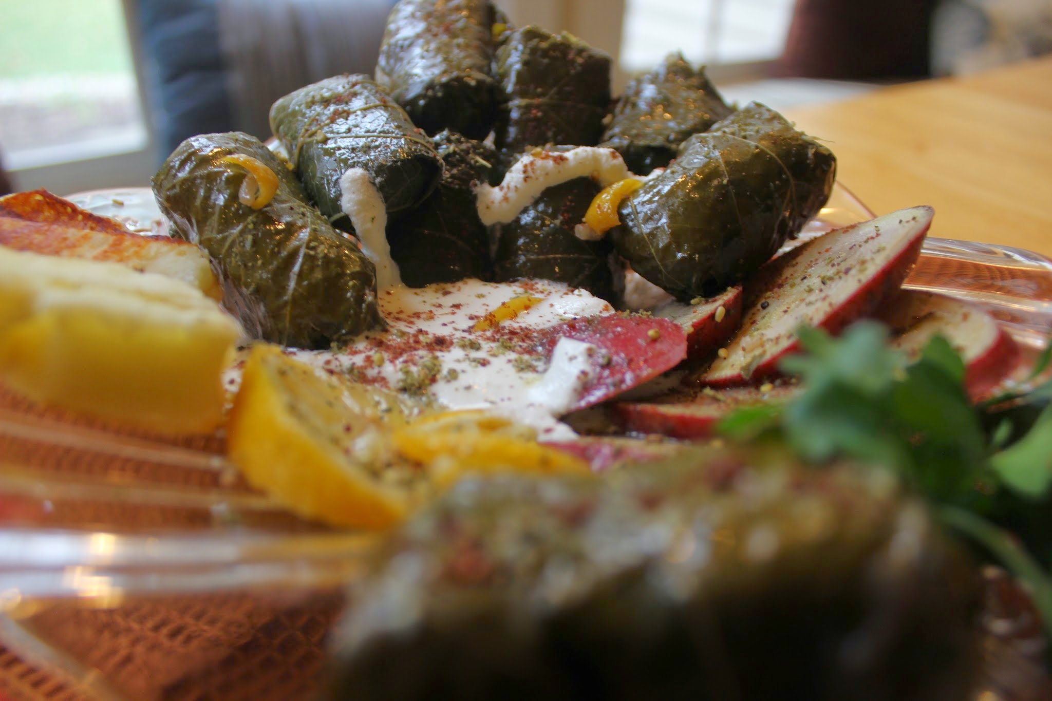
If you’ve ever snacked on delicious dolmas, then you know that the goodness of the grape vine goes beyond berries. Legend has it that the gods of Mount Olympus feasted on the tender leaves of the grape wrapped around morsels of rice or meat, alongside ambrosia and nectar! Although stuffed grape leaves are common around the Mediterranean, Greeks claim that dolmades were co-opted by the army of Alexander the Great to parcel out limited rations of meat during the seige of Thebes. Luckily, you only need to lay seige on your local Middle Eastern grocery store to find jarred leaves, preserved in brine. Unfurl them gently and give them a good wash to get started. It doesn’t hurt to have a glass of your favorite vintage, red or white, on hand before embarking on this project!