
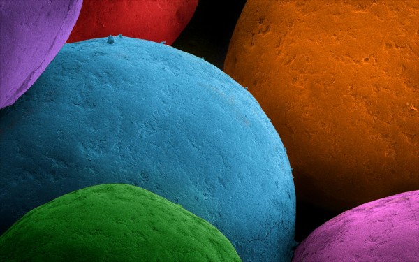
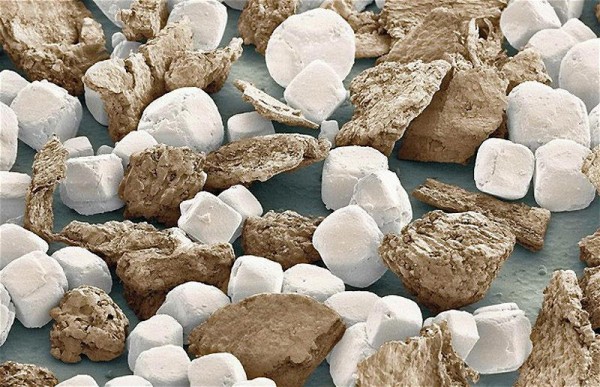
Under the Electron Microscope. WOW! Can you guess what these everyday objects are?
More: http://egotvonline.com/2012/03/13/25-everyday-objects-under-an-electron-microscope/


Under the Electron Microscope. WOW! Can you guess what these everyday objects are?
More: http://egotvonline.com/2012/03/13/25-everyday-objects-under-an-electron-microscope/

looks like colorful pasta
None of the above 😉 Think small! Much smaller!
hair, clothing fibers, pollen… and I don’t have a guess to those sort of ribbon looking things… hmm.
I cheated and looked at the descriptions of each photo. 😛
Absolutely amazing pics.
magnificent 🙂
I’ve got to look up what a sprinkle is. Where I live we don’t use them that much (fortunately).
No fun in guessing if you write the answer in the description 😉
细菌
Beautifull colors.
Nope, good guesses but…(answers in captions).
No need to dust, this is art. Right, Kimberly Brosnan ? 🙂
Sugar bits used to decorate cakes, Víktor Bautista i Roca .
Drew Sowersby , I think I know what you mean. Sometimes, it’s enough to just watch in awe.
you got some pollen on your polyester
LOL, Al Seaman !
Too brain dead to actually write up something on graph theory of neuronal branching, Feisal Kamil , which is what I really wanted to do this evening. So, eye candy to the rescue 🙂
Rajini Rao We’ll let this pass — but just this once, hear? < winks >
William McGarvey , by the time I even skimmed the paper in PLoS Computational Biology my neurons were all out of action potentials.
Rajini Rao That’s quite all right… we’re in a substantive area where I never had anything resembling an action potential.
Oh, I’m sure you’ve had a calcium spark or two light up those astrocytes, William McGarvey !
Besides, William McGarvey , I’ve seen those sparks in action 🙂
Rajini Rao Possibly so, but if wasn’t cast in psychometric jargon (preferably in covariance-structure models), I would never have known about it. (http://scholar.google.com/citations?hl=en&user=RZyrZXkAAAAJ).
well its another Under the Electron Microscope….beautiful….
nice
Wow – dust is so….beautiful.
Never seen something like this before so near.Inspiring ones
wats this rajini ?
I didn’t expect dust to look so clean. I expected it to be …. dustier.
some sort of smallest organic matter
Rajini Rao Photoshopped! Joking. When I used such a fancy tool there were no colours!
snow crystall under electron microscope!! is that possible?!!!!!!
Looked like avatar
The funny thing is that in output of electron microscope there’s no colors at all. I hope the author of program that paints different objects to different colors has much fun reading comments.
it is a good pattern
hiiiii
Alexander Tankeev You mean that color is not real? Disappointing.
must pick
This is so cool!
Prabat parmal yeah… I think even without decoration, it would have been real and interesting!
Thanks for fielding tech comments, Feisal Kamil , you’re a champ! R Prakash Prakash and Prabat parmal : False color or pseudo color is used in nearly all microscope images that I am aware. The digital signal output can be coded into any color your heart desires. The textures, shapes and other visual details are ‘real’ at least as the sample is prepared. I don’t think that a living object will survive electron microscopy, although there are variations that maintain more natural conditions such as cryo-EM where the sample is flash frozen.
They are inert until they enter a host cell where they can reproduce. You can also think of them as “obligate parasite”. The question of whether they are living or non-living is a pseudo controversy..depends on how you define life. Outside of the host, they are inanimate. But they “come alive” inside a cell. No different from a resting spore or seed, in my opinion.
Depends on whether they are structurally damaged after bombardment, I don’t know the answer. I do know of protein crystals that are later redissolved and shown to have enzymatic activity…this is done to show that the structure obtained from X-ray crystallography is that of an active conformation (=shape). Not a definitive argument, but it shows that the crystal survived exposure to rays. Certain cryoEM crystals must also survive imaging.
You’re welcome, keep those questions coming 🙂 What is your field by the way..I assumed you were an engineer, but perhaps you are studying one of the humanities?
Wow, such aesthetically beautiful mundane objects!!!
Point away, Feisal Kamil 🙂
Whoa!! Such brainstorming comments and discussions above!! great 🙂 count me in too 🙂 and keep them coming!
Also, ? And !
For the bemused, that translated to “keep the questions and amazement coming” 🙂
????????????
!!!!!!!!!!!!!!!!!!
🙂
Very good. The work is perfect!
Sorry I Cant guess.
Would you like to tell me about it?
Zia Anwar , click on each image..there is a caption in the comment box on the right.
thanks
Works or art.
I’ve seen some of these numerous times, yet I can only manage to remember the salt and pepper. I’m not so fond of being reminded of my faulty memory, but these kinds of images could flow through my stream three times a day and I wouldn’t mind in the least bit.
Crazy! The crack in steel is my favorite, looks like some kind of Martian landscape!
Mindbending! Rajini Rao Is this with some special aka high powered microscope or just the usual lab ones?
Oops: Ok so its an Electron microscope!
algie
great
Some kind of bacterial
http://viutube.lk/mobile-video-editing-සිංහලෙන්kinemaster-sinhala-review_v13619
viutube.lk – Mobile video editing සිංහලෙන්.Kinemaster sinhala review.
https://www.fivestuniversal.com/p/top-5-living.html
fivestuniversal.com – LIVING