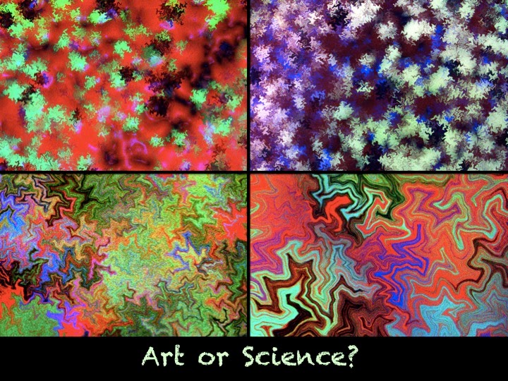
Art or Science?

Art or Science?

Are these pastel fractals the creation of an avant garde artist from some postmodern cubism movement?  You may be surprised to learn that these are high resolution images of bacterial populations growing on a petri dish!

Bacterial Art: First, the familiar E. coli bacteria were genetically marked with differently colored fluorescent proteins before mixing together on an agar plate. Each rod-shaped bacterium grows by division to give a single file of cells that is sensitive to small mechanical forces from neighboring cells pushing and jostling against each other. The line of cells buckles in a way that is predicted by fractal mathematics.  As the bacteria grow to form a confluent film, jagged boundaries emerge between differently colored clonal lines. Zooming in, the patterns are self-similar, repeating at scales from millimeters to micrometers! Mutant bacteria that form spherical cells don’t produce these fractal patterns. 

Form and Function: What do these beautiful images teach us? They help us understand how patterning happens on a nanoscale. In synthetic biology the goal is to engineer populations of cells to produce spatial patterns, synchronized signals and predictable behavior that can be simulated using simple, mathematically coded rules.  

Life Imitates Art? Oscar Wilde reversed the conventional when he claimed that life imitates art far more than art imitates life. What do you think he meant by this? It seems to me that this bacterial fractal “art” perfectly illustrates John Berger’s definition of Cubism: “The metaphorical model of Cubism is the diagram: The diagram being a visible symbolic representation of invisible processes, forces, structures.”

Reference (and more beautiful images): http://data.plantsci.cam.ac.uk/Haseloff/resources/LabPapers/Rudge2013.pdf

  #ScienceSunday  

Posted in Rajini Rao | Tagged | 92 Comments

Fixing a Hole: Better (Spider) Web Design

Fixing a Hole: Better (Spider) Web Design

⎈ From tiny webs like the one “repairing” a hole in a leaf seen in the image, to giant orbs spanning 25 meters across rivers and lakes, the architecture of spider webs can teach us a thing or two about engineering. After all, spiders have been spinning silk for 400 milion years and now number at least 41,000 species spread out over every continent, including Antarctica. Each spider produces many different types of silk covering a range of mechanical properties: from the steely dragline silk in the radial strands to sticky capture silk that forms concentric circles in the web. Yet, only few spider silks have been studied, mostly at random, sometimes simply from the researcher’s own backyard! 

Bioprospecting: By combining fields as diverse as natural history, ecology, taxonomy, behavior and biomaterial science, researchers found that the Darwin’s Bark Spider (Caerostris darwini), a giant Malagasy riverine orb-weaving spider, produces the toughest silk discovered to date. Outperforming steel and Kevlar, the radial web threads of this spider have unusual elasticity, absorbing more kinetic energy upon prey impact so that they stretch, instead of fracturing. This allows the spiders to occupy a new ecological niche- the flyways above rivers where they can catch unsuspecting insects and even small birds and bats. Don’t you agree that scientists should get out of their labs and explore new habitats as well?!

Biomimicry: In nature, tiny amounts of metals penetrate protein structures to change their properties. These “impurities” are found in jaws, claws and cuticles where they impart additional toughness to biological material. Inspired by nature, scientists purposefully introduced zinc, titanium or aluminum into spider dragline silks by using a multiple pulsed vapor-phase infiltration method. The resulting material was tougher and more stable to environmental damage. Now this is the stuff of Spider Man!    

Free Reads: New Opportunities for an Ancient Material (2010) Ometto and Kaplan. Science. http://www.ncbi.nlm.nih.gov/pmc/articles/PMC3136811/

Bioprospecting Finds the Toughest Biological Material: Extraordinary Silk from a Giant Riverine Orb Spider (2010). Agnarsson et al. PLOS ONE http://goo.gl/CcSMTd

The Beatles-Fixing a Hole: https://www.youtube.com/watch?v=j0I2ZrBuFdQ

Photo Credit: Bertrand Kulik 

#ScienceSunday  

Posted in Rajini Rao | Tagged | 49 Comments

How to Boil Water

How to Boil Water

❉ In breaking news, scientists have figured out how to boil water – at least 3 times more efficiently and producing twice as much steam. Before you shake your fist at “wasteful research spending”, this isn’t really about your whistling tea kettle! 

❉ Phase change heat transfer processes (boiling, condensation) are a big part of everyday technology from water purification and HVAC units, power plants and cooling electronics.   When water boils, a thin layer of steam can coat the heated surface, insulating it and drastically cutting down on the efficient transfer of heat to liquid. This can lead to surface burnout and a destructive condition known as critical heat flux. What is needed is a surface that discourages the vapor from sticking and wicks in water to quickly re-wet the heated surface. To create a superhydrophilic wicking surface, Drexel University scientist Matthew McCarthy turned to biotemplating with….viruses! 

❉ The tobacco mosaic virus causes mottling of tobacco leaves, as its name implies, but is harmless to humans. It was the first virus ever to be discovered (in the late 1880’s) and is constructed simply of repeating units of a coat protein, wrapped around a single, helical strand of genetic material (RNA). A few tobacco plants can produce billions of virus particles, so it’s cheap to make. Dr. McCarthy tweaked the coat protein so it sticks to any engineered surface- from silicon to steel. After dunking the surface in a viral broth, nickel and palladium are added to grow a metallic grass

❉ The viral tendrils work like a wicking surface, drawing down water to replace what’s boiled away.  It’s the same idea behind thermal fabrics designed for athletes which draws moisture away from the body. They say a watched pot never boils. I’d volunteer to test a virally coated tea kettle, how about you? 

Waterproofin’ with Hydrophobin: This old post shows how a fungal spore protein can do the opposite, creating a superhydrophobic surface that repels water but allows gases to exchange. 

https://plus.google.com/u/0/+RajiniRao/posts/bf9gVFkaTxQ

News Story and Short Video: http://drexel.edu/now/archive/2015/March/TMV-heat-transfer/

Ref: M.M. Rahman, E. Ölçeroğlu, and M. McCarthy, “The Role of Wickability on the Critical Heat Flux of Structured Superhydrophilic Surfaces”, Langmuir 2014, 30 (37), pp 11225–11234.

#ScienceSunday  

Posted in Rajini Rao | Tagged | 101 Comments

Cleo Gets a Coat!

This tortoise is wearing a 3D printed shell. Why?

⎈ Cleopatra is a teenager who seems pretty happy with her bright, new red coat. What’s unusual is that Cleo is a leopard tortoise! Also, her coat was 3D printed from a corn-based polylactate polymer by student designers at Colorado Technical University. No, this isn’t a new challenge on Project Runway, although the coat does stylishly drape over Cleo’s shell. It’s actually a 600 hour labor of love that will hopefully let Cleo enjoy a long (80+ years), tortoise life. 

⎈ Tortoises in the wild have smooth and convex shells. Unfortunately, when bred in captivity, tortoise shells grow in raised bumps known as pyramiding. When Cleo horsed around with her tortoise friends, the deformed shell wore through in spots, making her susceptible to bacteria and other infections. Fortunately, her red coat is temporary and her shell is expected to heal in a few years. 

⎈ Scientists are not sure what causes pyramiding. Too much dietary protein (Cleo is a herbivore) is one culprit. Not enough bone calcium is another. One study showed that raising the humidity helped. Until we solve the problem or stop breeding tortoises in captivity, we have prosthetics -not just for humans, but for our four legged friends too. 

News Story: http://goo.gl/qtZw7V

Ref: Influence of environmental humidity and dietary protein on pyramidal growth of carapaces in African spurred tortoises (Geochelone sulcata) (2003). C. S. Wiesner andC. Iben. Journal of Animal Physiology and Animal Nutrition. Volume 87, pages 66–74

Posted in Rajini Rao | 67 Comments

What Autism can Teach us about Brain Cancer

What Autism can Teach us about Brain Cancer

Glioblastoma multiforme, or GBM, is a deadly cancer with median survival of only 12-15 months. Recently, we found a gene that had been previously implicated in autism to also contribute to GBM. The gene NHE9 makes a protein that exchanges sodium ions for hydrogen ions (also called protons) across the boundaries of endosomes, hence it’s moniker “sodium-hydrogen exchanger”. But what are endosomes and why is the function of NHE9 important?

Highway Traffic: All cells contain many “cargo packages” surrounded by membranes, depicted in the expanded view as a blue compartment in the figure below. These so-called endosomes carry newly minted proteins to specific destinations throughout the cell and haul away old proteins for destruction. Key to their “shipping speed” is the level of acidity inside the endosomes. Acidity relates to the number of protons, which are controlled by balancing the activity of “pumps” that push protons into endosomes to increase their acidity with that of “leaks,” like the protein NHE9, that remove protons. 

There’s a Hole in the Bucket: You can think of endosomes as leaky buckets of water. Altering either the faucet or the leak rate can dramatically change the water level in the bucket. In autism, NHE9 is mutated and non-functional. In the absence of proton leak, the endosomes become too acidic and prematurely clear away important proteins on nerve ends, leading to neurological dysfunction. Helper cells called astrocytes cannot clear away neurotransmitter signals fast enough, and this leads to hyperexcitability or seizures associated with autism.

Too Much of a Good Thing: in contrast to autism, NHE9 is overactive in brain cancer, causing endosomes to leak too many protons and become too alkaline. This slows down the “shipping rate” of cancer-promoting cargo and leaves them on the cell surface for too long where they inappropriately prolong signals of growth and migration, the two main characteristics of invasive cancer cells. Fortunately, when the leak is plugged by inhibiting NHE9 with drugs, tumor growth is blocked. Currently, the drugs are not good enough to use on patients, so an important step going forward will be to discover better drugs that target NHE9. These could be used in combination with other drugs for treatment of this deadly disease. 

Paper: A leak pathway for luminal protons in endosomes drives oncogenic signalling in glioblastoma. Kondapalli et al. (2015) Nature Communications http://goo.gl/dAa5NG

Johns Hopkins News Story: http://goo.gl/XAsGDb

A Part of the Puzzle: NHE9 and Autism https://plus.google.com/u/0/+RajiniRao/posts/fsNzo1yKsQG

  #ScienceSunday  

Posted in Rajini Rao | Tagged | 34 Comments

Ooh, Lab Bench Cake!

Ooh, Lab Bench Cake!

Who can guess what all the items are? Bonus points if you tell us what they are used for. #showoffyournerdyside  

Via http://www.cakecentral.com/gallery/i/1929656/lab-bench-cake

#ScienceEveryday  

Posted in Rajini Rao | Tagged , | 95 Comments

Souring on Sweeteners: Why Diet Sugars Don’t Work

Souring on Sweeteners: Why Diet Sugars Don’t Work

Sweet Serendipity: It was 1878, when a beaker of coal tar compounds boiled over in the chemistry laboratory of Ira Remsen at the newly founded Johns Hopkins University. Researcher Constantine Fahlberg cleaned up the mess, but later at dinner, his hands tasted surprisingly sweet as he put a piece of bread in his mouth. And this is how the first artificial sweetener was discovered! Named saccharin, it was 300 times sweeter than sugar. Soon, it was being prescribed to President Theodore Roosevelt, to counter his corpulence. More low-calorie sweeteners followed: sucralose, stevia and neotame, the last one being 10,000 times sweeter than table sugar. Today, 30% of adults and 15% of children in the U.S. consume low calorie sweeteners. A sweet deal, right?

Caloric Contradictions: Unfortunately, counterintuitive to expectations, studies show that people who consume large amounts of artificially sweetened drinks gain more weight and body fat compared to those who don’t. Could this be a case of reverse causation? Perhaps, increased body weight encourages people to turn to non-caloric sweeteners. However, this has been ruled out by (i) controlling for baseline body weight at the start of the study and by (ii) looking at weight changes in people who are not overweight to begin with. Another possibility is cognitive distortion: because non-caloric sweeteners are perceived to be healthy, we take that as permission to consume more high-calorie foods. Imaging studies of the human brain reveal a metabolic cause: unlike ordinary sugar, non-caloric sweeteners do not trigger the reward circuits that initiate satiety and fail to activate normal pathways of insulin release needed to deal with caloric loads. 

Diet to Diabetes: New research in both mice and humans showed that artificial sweeteners also change our gut microbiome, leading to glucose intolerance, the first step to diabetes. Surprisingly, if the feces from saccharin-fed mice was transplanted into mice whose guts were first cleared of bacteria by antibiotics, the sugar handling defect could be induced in the healthy mice. Oh, expletive!

The Archies sang, ♫ Oh honey, sugar, sugar..you are my candy girl and you’ve got me wanting you

https://www.youtube.com/watch?v=0MiQzAo6Cp8

REF: Artificial sweeteners produce the counterintuitive effect of inducing metabolic derangements. Susanne Swithers (2013) http://www.ncbi.nlm.nih.gov/pmc/articles/PMC3772345/

News Story & Link to Nature paper: http://www.nature.com/news/sugar-substitutes-linked-to-obesity-1.15938

  #ScienceSunday  

Posted in Rajini Rao | Tagged | 119 Comments

Cancer: What’s Luck Got To Do With It?

Cancer: What’s Luck Got To Do With It?

Grandfather smoked like a chimney, ate bacon everyday and lived to be 90. Yet, your best friend, a lifetime vegan who exercised regularly, succumbed to breast cancer in her thirties. We’ve all used these anecdotes to try to make sense of the deadly scourge that is cancer. So do this week’s headlines in the popular press, Biological bad luck blamed in two thirds of cancer cases give us license to bring on the bacon and booze? Not so fast, as the study just published in Science by Johns Hopkins researchers Cristian Tomasetti and Bert Vogelstein was much more nuanced than the headlines suggest.

♦ The premise of the study was the puzzling observation that some tissues give rise to cancers a million times more frequently than others: for example, the lifetime risk of being diagnosed with cancer is 6.9% for lung, 1.08% for thyroid, 0.6% for brain, 0.003% for pelvic bone and 0.00072% for laryngeal cartilage. Even within the digestive tract, cancers of the colon (4.8%) are much more common than stomach (0.86%) despite both tissues being exposed to the same carcinogens and dietary insults. To make sense of this, the researchers turned to cancer stem cell theory: that malignancy is caused by mutations in a small number of stem cells that retain a lifetime ability to divide. They then searched the literature to estimate the number of stem cells in each tissue. What they found was that tissues with higher populations of stem cells were more prone to cancer. This linear correlation (R=0.8; R^2=0.65) was pretty good, extended over 5 orders of magnitude (see graph), and makes sense since we already know that every time DNA replicates there is a finite chance of making errors and that the more mutations in DNA the greater the chance of some of them triggering cancer. In simple terms, a large part (estimated two-thirds; see R^2) of the variation in cancer risk between tissues is due to the difference in their stem cell population. This does not translate into “two-thirds of an individual’s risk of cancer is due to dumb luck”!   

Environmental, lifestyle and genetic risk factors pile on top of the basic risk of random mutations from stem cell divisions. To identify cancer types (red circles) in which the contribution of environmental and inherited factors was especially high relative to the random DNA replication-driven component, researchers used an unbiased clustering algorithm that used the product of the log values of the x- and y-axes in the graph below. What they found was consistent with what we already know about some cancers. Smoking greatly increases risk of lung cancer by ~18-fold for both sexes (23-times in men, 13-times in women), as seen by the higher risk incidence of lung cancer for smokers compared to non-smokers in the chart. People with familial mutation in the APC gene have a 100% rate of colorectal cancer unless the colon is removed. Infection with Hepatitis C increases risk of liver cancer by 10-fold. 

♦ This new analysis explains some puzzling facts: the same APC mutation has a much higher chance of giving rise to colon cancer instead of duodenal cancer because there are 150 times more stem cell divisions in the former compared to the latter. Another example is that basal epidermal cells and pigment cells of the skin (melanocytes) are exposed to the same carcinogen (UV radiation) at the identical dose. Yet, basal cell carcinomas are much more common than melanomas. The authors argue that this is explained by the underlying difference in rates of stem cell division. 

Why should we care about this analysis? Understanding the underlying risks contributing to each cancer type should determine the best public health strategy to tackle it. Early detection should be the main focus for prevention of cancer types largely driven by random errors in DNA replication, whereas vaccines against infectious agents or altered lifestyle will be key to reducing incidence in cancers with high environmental risk.  

REF: http://www.sciencemag.org/content/347/6217/78.abstract

Inset image: http://www.sciencephoto.com/media/254142/view

#ScienceSunday   #cancer  

Posted in Rajini Rao | Tagged , | 101 Comments

Champagne Science

Champagne Science

If an estimated 360 million glasses of champagne will be toasted this New Year’s Eve, how many bubbles would they release? To figure this fun fact, we’ve got to get back to basics. 

It’s a Gas: In 1810, French chemist Joseph-Louis Gay Lussac determined that in fermentation, glucose is converted to equal parts of ethanol and carbon dioxide gas according to the equation: 

                   C6H12O6 > 2C2H5OH + 2CO2

To make champagne, this basic wine is dosed again with glucose (typically 24 g/L) for a second round of fermentation, yielding 11.8 g/L of CO2. All that CO2 is dissolved, under pressure (as much as 90 psi), inside the champagne bottle. 

Don’t Shoot Your Eye Out!: The American Assoc. of Ophthalmologists warn that a champagne cork can launch at 50 mph! Why is this? Henry’s Law (1803), paraphrased, says that the amount of gas dissolved in a liquid is proportional to the pressure of that gas above the liquid. When a champagne bottle is uncorked, the CO2 in the space above the liquid escapes, forcing the dissolved gas to come to a new equilibrium. This results in release of about 5L CO2 per bottle. 

Fizzy Physics: Dr. Gérard Liger-Belair didn’t care for the over-blown bubble estimates being bandied around the popular press. So, armed with plenty of free samples from Champagne Houses Pommery, and Veuve Clicquot Ponsardin, he buckled down for some serious science (it’s a hard life for a noble cause, hic!). After considering such factors as the van’t Hoff equation for temperature dependence,  the critical radius for bubble nucleation and ascending bubble dynamics, he published his findings in a recent issue of the Journal of Physical Chemistry. The answer to our question? If 100 ml of champagne is poured straight down the center of a vertically oriented crystal flute, about one million bubbles will form, “if you resist drinking from your flute”.  But, who’s resisting? 🙂

With that, I raise my glass to yours along with approximately 360 trillion other bubbles world wide, to wish you a Happy New Year! 

REF: How many bubbles in your glass of bubbly? (2014) Gérard Liger-Belair http://pubs.acs.org/doi/abs/10.1021/jp500295e

Pop Sci: Back story on champagne research via Chad Haney http://www.rsc.org/chemistryworld/2012/05/raising-glass-champagne

      #ScienceEveryday   #HappyNewYear   

Posted in Rajini Rao | Tagged , | 124 Comments

A Very Marine Xmas!

A Very Marine Xmas!

At the very bottom of the marine web of life, are tiny plankton that drift seemingly aimlessly with the ocean’s currents. Phytoplankton are responsible for 50% of the earth’s photosynthesis, and fix a 100 million tons of CO2 each day. Remarkably, every 2-6 days, the entire biomass of plankton is consumed by filter feeders, from barnacles to baleen whales. Did you know that the distinctive smell of “sea air” is from chemicals given off by decomposing phytoplankton? 

Micro Marvels: In homage to these remarkable creatures, scientist Dr. Richard Kirby made this composite Christmas card entirely from plankton: paddle worms, sea angels, protozoa and jellyfish (read more here: http://goo.gl/GAv5lG). 

Delightful Doggerel: My Christmas jingle is just for fun, inspired by Chemistry Carols from the Wesleyan University page (http://goo.gl/QKTjfX). More fun Lab Carols from the staff of ASBMB can be found here: http://goo.gl/rkxW7s .

Over to You: Go ahead, be a sport. Share your science-y holiday verse here 🙂

#ScienceEveryday   #merrychristmas  


Posted in Rajini Rao | Tagged , | 57 Comments