
The Unwanted Guest: Parasites are clever about overstaying their welcome, co-evolving with their unwilling hosts to survive and propagate. Viruses, for example, insert their DNA into the host chromosome, ensuring that they are passed on to the daughter cells.
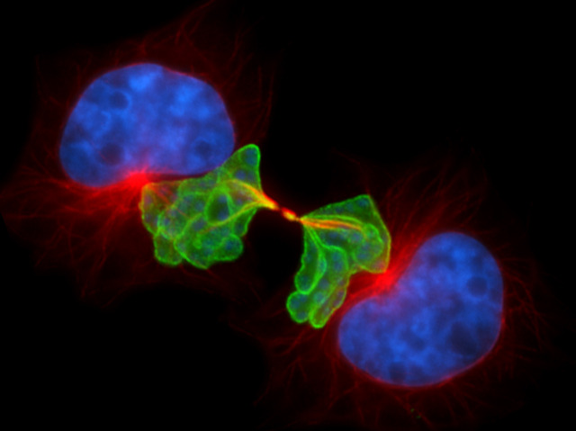
This clever protozoan, Theileria (in green), aligns itself with the mitotic spindle (shown in red) as if it were a chromosome. When the spindle of the dividing host cell pulls apart to form two nuclei (in blue), the parasite goes along for the ride. Theileria infects cattle via tick bites and resides in white blood cells. Other disease causing protozoan parasites like Plasmodium and Toxoplasma also co-opt microtubules to help in cell invasion. This new finding means that drugs that target microtubules could block parasite spread.
Free Read: http://goo.gl/NM4uv
Image from Dirk Dobbelaere, University of Bern (Switzerland) via BPoD.
#sciencesunday

ZOMG, that is seriously creepy! I think Buddhini Samarasinghe has posted on zombie parasites before.
Here they are: zombie roaches! http://goo.gl/JYDK8
Happy ending, as Buddhini says, “the only good cockroach is a dead cockroach” 🙂
It’s all for science, Do it! 😀
Beautiful!
As often happens, it’s the image that captures the imagination first, Malin Christersson 🙂
Clever Theileria! It seems awfully tiny for a protozoan (size of a chromosome?), so I did an image search on google and found the most interesting images, mostly of ticks, and one of a chromosome being hijacked – “The New Scientist” http://www.the-scientist.com/?articles.view/articleNo/30719/title/Mitotic-Hijacker/
G+ is all about visualization, which sometimes is unfortunate in my humble opinion. 😀
That’s a great read, thanks for the link BiologyCorner ! It was nice to get the author’s perspective on the work.
Theileria is described as a syncytium in this stage of its life cycle..sort of amoeba like, I imagine. It’s interesting that it does not interfere with chromosome segregation, or host cell mitosis.
hello how r u rajni 09907421220 coll plz i mis you
LOL, Feisal Kamil , I’m afraid to try it 🙂
And thanks, you’re a great pal.
Raj, I’ll help you out, here’s a toll free number that will connect you. 1-888-642-6257
Rajini Rao you post is called unwanted guest hehe.
It was an unwitting invitation, wasn’t it Chad Haney ? 😀
I think we have dislodged his invasion, though, thanks for your help!
Cool post! :D)
Great post Rajini Rao
Very nice post!
Lacerant Plainer gmta !
Great post, and always such awesome wit amongst the scientists, :).
Hehe yep Euro Maestro 🙂 I see our comments were a minute apart, but it took time for yours to be visible to me.
That is fascinating; I love science stuff like this. An to think we still only know about 70 percent (if that) of what the small world has to offer.
how this is imaged and observed ?? what techniqued
?
pinto xavier , this is standard immunofluorescence microscopy using antibodies and DAPI for the nuclear stain. They used confocal imaging, where fluorescence is collected from a thin optical section to make the image sharper. The details are in the methods section of the PLoS Biology paper in the link that I provided.
The image looks awesome. I guess the host does not think so.
Does this imply common, already investigated drugs, such as colchicine?
Yes, I agree. Several anti-cancer drugs target microtubules, including colchicine and vinblastine.