
The One Ring: Structure of the c Ring of the Proton Pumping ATP synthase.
Protons are pumped across membranes to drive a rotary shaft that in turn makes ATP, the “currency of life”. ATP is used to fuel virtually all energy requiring processed in our body. For a beautiful animation of this rotary pump, see: http://goo.gl/gjiZN
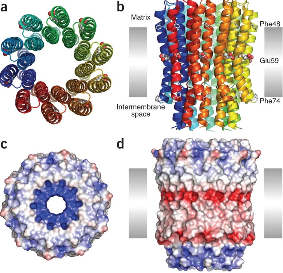
Protons are ferried by the the c ring (image a), as in a merry go round. Each subunit carries a proton binding site (Glu59, image b), as it rotates. Images c and d show the electrostatic surface of this rotor, with acidic charges in red, basic in blue and neutral as white.
This structure is hot off the press and was presented by my friend David at the ATPase conference I am attending in Aspen, Colorado. It was obtained by making crystals and imaging with X-rays.
Ref: Structure of the c10 ring of the yeast mitochondrial ATP synthase in the open conformation. Jindrich Symersky, Vijayakanth Pagadala, Daniel Osowski, Alexander Krah, Thomas Meier, José D Faraldo-Gómez & David M Mueller ; Nature Structural & Molecular Biology 19, 485–491 (2012) doi:10.1038/nsmb.2284
David’s Homepage: http://web.mac.com/mueller.david/iWeb/LMB/dm_index.html

Protons as in the subatomic proton?
Amazing!
Shah Auckburaully , yes, exactly that. Protons as in the hydrogen ion.
Cells impress me.
Me too, Shah Auckburaully 🙂
You rock, Rajini Rao !!
Super cool Rajini Rao as always 🙂
Wanted to ask, how is the ATPase actually isolated and crystallized?
Protons are pumped across membranes to drive a rotary shaft that in turn makes ATP, the “currency of life” and Rajini Rao good post pump G+, which is “currency of life for G+”.
Deeksha Tare , the ATPase is isolated by good old fashioned biochemistry..starting with many liters of cells (60 liter batches!), breaking them and isolating proteins by differential fractionation, centrifugation, etc.
Beautiful. It’s amazing that he was able to get that resolution: 2 angstroms is just 4 times the size of hydrogen so substantially less than the size of one residue. (I’ve seen electron diffraction images and they just look like noise!) It appears that 10 subunits make up the whole assembly. Does that mean it consists of 10 identical protein molecules or are a variety of proteins involved? And is the c-ring a separate protein or a unit of a larger protein? (Thanks, sorry to ask so many questions!)
The ATP Synthase Enzyme
This is one of my favorite animations of ATP synthase that I show to my intro biology classes. Interestingly, its made by a creation science organization and they use it as an argument for intelligent design. However, their argument has a big, obvious mistake that my intro biology students always catch. See if you can catch it too!
Rajini Rao tell your crystallography pals to keep an eye on the next NIH Common Fund announcement. http://goo.gl/afwM1 At the Common Fund workshop I attended, the group that I was in liked a proposal to fund projects to create tools to help capture dynamic events of proteins at sub-microscopic level (protein-protein interactions, allosteric processes). They mentioned something about a laser source, but it’s not my area and apparently I’m not so good at taking notes.
Nice. I remember that awesome animation from your earlier post. Yeas ago I was particularly interested in this coenzyme when I was thinking of supplementing with Creatine Monohydrate. ATP -> ADP + (P)Cr -> ATP was a big win!
As a avid Scuba/Rebreather diver. I really wanted to get to the bottom (atomic) of CNS O2 exposure. So I went on Google and took a look at how 1.6 partial pressure of O2 impacts cellular exchange. From what I gather, cells are like Jet Engines, too little O2 blackout conditions where there isn’t enough energy to drive functions. too much O2 and the JET engine overheats with runaway RNA/DNA synthesis leading to convulsions.
Matt Kuenzel , I’m delighted to answer your questions. This is X-ray diffraction crystallography and the resolution is determined by the quality of the crystals..good crystals diffract further, providing more atomic level information. Electron microscopy should in theory also give atomic resolution but in practice the electron beam can destroy the biological sample so that the images are purposefully underexposed and overdeveloped..making them grainy. I was planning to post on EM methods along with a beautiful EM image, hopefully soon.
As for the c ring, in this case it is a ring of 10 identical proteins (subunits). In some organisms it could be 12 or 13 or more. Each subunit has two helices (20 can be counted in the image)..only one has the proton binding amino acid side chain (acidic glutamate). There are other proteins, not shown in this preparation. There is one large subunit (“a”) that provides a positive charge or counter ion that helps knock off the proton. This subunit makes two hemichannels so the protons can’t just leak across the membrane. Instead, they enter through one half channel, ride around the ring and exit through the other half channel, across the membrane!
Eric Scott , please do not show your class the animation you linked to: it is not a faithful atomic representation at all! The link in my post is from Dr. John Walker’s homepage http://goo.gl/gjiZN …he is the one who solved the structure and won the Nobel for it. Also, try this version which is old but textbook quality: ATP Synthase
The ATP synthase is actually a terrific example of evolution in action, both at the level of individual parts and as a whole. In fact, I teach my class evolutionary aspects of it to better appreciate how it works. In archaebacteria, there is an ancient version that functions as an ATP synthase. In eubacteria, there is a slightly different version that shares origin with our mitochondrial version (illustrating the endosymbiotic origin of mitochondria). Another derivative lost ability to synthesize ATP and works only in the opposite direction (ATP-dependent proton pump). This is the vacuolar or V-ATPase.
At the level of individual subunits, there is clear evidence for evolution as relating to function. These creationists don’t know much so they can spout nonsense. As the saying goes..a little bit of knowledge is a dangerous thing!
So the other animation is not structurally correct? I mostly liked it for the quality of the graphics, and as a quiz about what they get wrong in that video. I’ve also shown the animation from the link in your post in the past as well.
Eric Scott , if you look at it, the model is just a bunch of random balls stuck together in the general shape of the ATP synthase. It does not represent any protein at all. Compare it to the link I posted or even to the c ring space filling model and you will see that the latter are real proteins. I saw this one too, which shows just the ATP synthesis step Rotation of the gamma subunit effects ATP synthesis Let me know if this makes sense!
I am just a raw amateur on the subject. I know, in case my memory from High School is still working right, that ATP is Adenosin TriPhosfate, and the fuel of the miopower by mean of Krebs Cycle.
My question is: The rotation of F1 should increase to answer the miopower demand when the muscles are under extreme exigence, or it has an average level which is mostly the same under all circumstances?
Thanks for showing fantastic knowledge.
Aww thanks, Rashid Moore 🙂
Alfonso Ramirez , your memory is fine! ATP is indeed adenosine triphosphate. When it breaks down to ADP (adenosine diphosphate), energy is released for all sorts of jobs the cell needs to do. The rotation rate of F1 depends on the proton gradient, which in turn depends on the metabolism of the mitochondria (Krebs cycle and the electron transport chain), which is regulated by the needs of our muscle. I’ll ask around at my conference if someone knows of a more direct way that the rotation is controlled by the energy needs of the muscle, okay? Cheers!
We need a Cellular Biology Hangout on Air. Fascinate us. Christiane Cantin , fancy joining Rajini Rao into an informative topic discussion on Air?
I see now. Thanks!
Thanks, Rajini Rao how they coaxe these bio- molecules to line up for X-ray crystallography is a mysterious art!
Matt Kuenzel , crystallizing membrane proteins is particularly difficult..close to impossible, but fortunately for us, not quite. I saw one structure that was determined from a single diffracting crystal..out of >60,000 trials. Others have worked on crystallizing the same membrane proteins for decades..there is a black art and lots of luck involved in this field 🙂
Worth noting that several Nobels have been given out..for the first membrane protein and first ion pump and first ion channel structure solved by this method.
Wow, all this for a proton pump post …plenty more ion pumps where that came from 😛
What response was that Grant Burke 😉
LOL Mahesh Sreekandath & Feisal Kamil ! Good morning and good night!
Oh, no problem, you can check them later on 🙂
Thank you for this post Rajini Rao
This is one circle that everyone is plussed in. Or dead.
Why do we have to carry such heavyweight words in Biology? I feel so overburdened.
Haha Mahesh Sreekandath ! It’s been so long since someone called me by that name! Thanks for reminding! 🙂
This is mindboggling in the best, most constructive sense of that word.
Thanks for the answer, Rajini.I do not dare to give ideas on the amazing technical achievements of you.I think it should be a limit of the rotation, because of the medium. But may be the pressure exerted by the pressured muscle can be read,and transmitted to all systems compromised in the task of powering muscles. I have been a First Division Rugby player,and also climbed high mountains,and metabolic system was always of interest to me.