
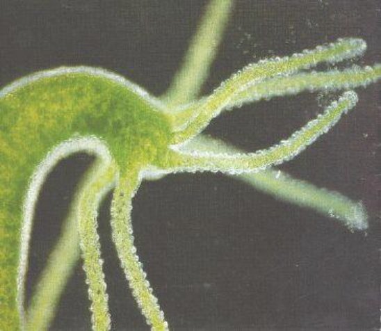
Hydra Vision: Implications for Evolution of the Eye. The freshwater polyp, Hydra (a relative of jellyfish) catches prey with a potent sting from its waving tentacles. The tips of these tentacles were known to be sensitive to touch and to chemicals, and now, to light. But the Hydra has no eye. Or does it?
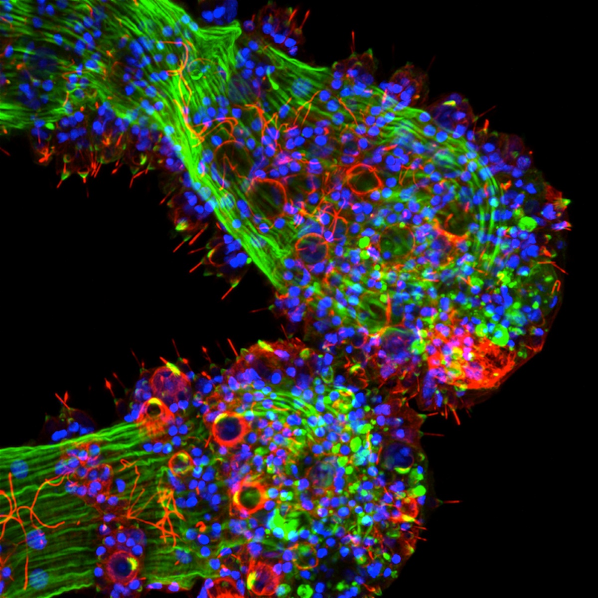
• Researchers found the basic components of our visual signaling mechanism in the tentacle cells (Image 2): the photosensitive protein opsin, the cyclic-nucleotide-gated (CNG) ion channel, and arrestin (a protein that shuts off opsin). They then showed that firing of the stinging cells (cnidocytes) was inhibited by bright light, and that a drug that blocked the CNG channel reversed this light inhibition (Image 3). Possible functions of light sensitivity could be to regulate feeding patterns or to sense shadows cast by approaching prey.
• This means that the biochemical pathway central to vision in our highly complex eye is found in this boneless, brainless animal. Studying primitive “vision” is like looking back in time to the early steps in evolution that predate the development of our eyes .
REF: http://www.biomedcentral.com/content/pdf/1741-7007-10-17.pdf


Eye see what you did there.
After that terrible joke, I’m going to get some eyece cream.
Gasp, you mean it’s not ‘god-did-it’?! Great post Rajini Rao as always 🙂
Oh, and I made this for you. Consider yourself warned 😀 http://i.imgur.com/LDhmg.jpg
Thanks, Buddhini Samarasinghe ! I’ll keep an eye out for them 😛
Fascinating, Rajini Rao. Thanks for sharing, and the excellent summary.
Truly amazing. Thanks for this….a must share!
I like the idea of using gelatine-dipped fishing line to collect harpoons.
Deep inside the paper I see this (chapter “Methods”, section “Animal culture and cnidocyte capture assay”): “For cnidocyte capture trials, we placed healthy and responsive animals in either dim blue light (0.1 W/cm^2) or bright blue light (2.8 W/cm^2) for 6 h prior to probing.”
Now, 0.1 W/cm^2 (“dim light”) is 1kW/m^2, i.e. 2/3 of sunlight from the zenith (integrated over the whole spectrum). The thing called “bright light” is 28 kW/m^2 and would heat an irradiated body to about 800 Kelvin by Stefan-Boltzmann’s law (unless well cooled). Either I am misunderstanding something very badly, or the hydras were cooked, or they were in a thoroughly flushed aquarium[0]. Any ideas?
[0] 2.8 Watts will heat a cm^3 of water by ca. 0.7 Kelvin per second, i.e. at 2.8 W/cm^2 and an 1 cm thick water layer we need a flow of at least 0.7 cm/s to limit the temperature change to 1 Kelvin.
PS: The paper describes that the light intensity was measured, therefore I’d exclude the possibility of confusing light output power and consumed electrical power (i.e. neglecting the non-perfect efficiency of LEDs).
This is truly a find “biochemical pathway central to vision in our highly complex eye is found in this boneless, brainless animal. Studying primitive “vision” is like looking back in time to the early steps in evolution that predate the development of our eyes” that research is being done in this area!
Ralf Muschall , I can email the lead author with your question and let you know the response. Meanwhile, the fact that bright light suppression was blocked by 1 micromolar cis-diltiazem means that the hydras were not cooked or harmed by the light. (If the lower response in bright light was due to dying Hydras then of course no drug could rescue them.) The Methods state that the LED array was placed at varying distances from the Hydras to adjust brightness. My understanding is that the labels under the bar graph refer to the power intensity of the source but not what the animals are exposed to.
Ralf Muschall — I suspect that while the light does have the intensity you’ve calculated, the actual die size of the LED was less than a couple square millimeters, meaning the total emitted power was more like a few watts.
This is like art in nature, I have to learn this staining techniques seems like a lot of fun 🙂
kiran kumar , not precisely “stains” but immunofluorescence. You would need a pricey laser scanning confocal imaging microscope 🙂