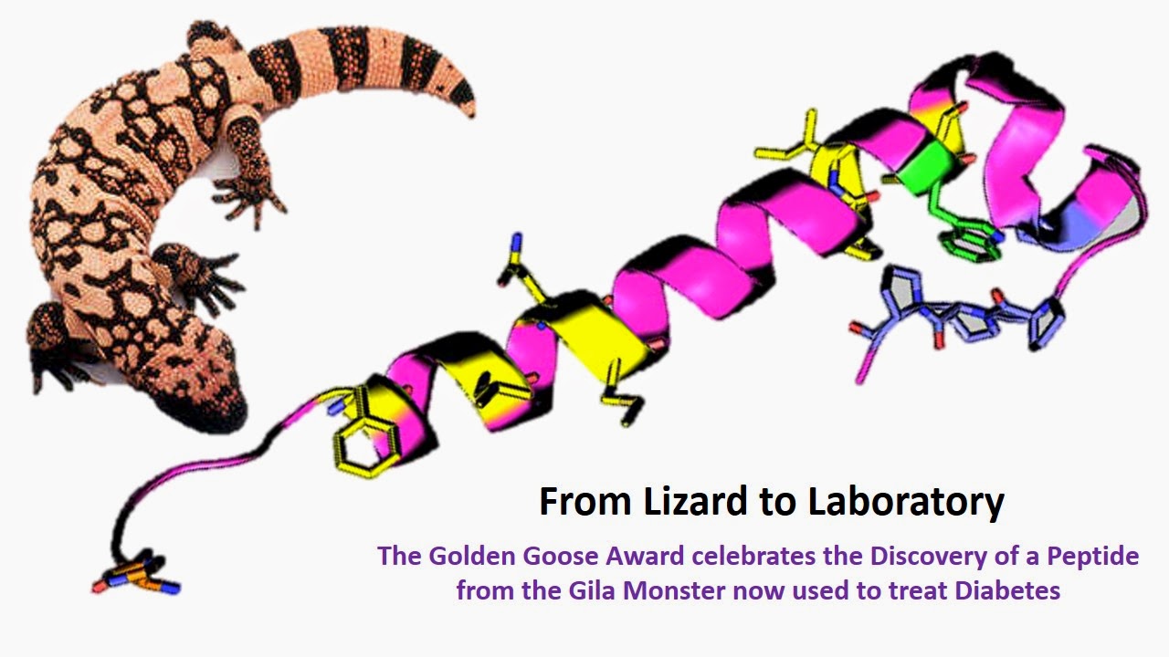
The Golden Goose Award celebrates federally funded research that is seemingly obscure but turns out to have an unforeseen positive impact on society. This year, the prize goes to Dr. John Eng whose discovery of the peptide exendin-4 from the venom of the 2-foot long pink-and-black Gila Monster has provided relief to millions of diabetics.
From Lizard to Laboratory: In 1990, Dr. Eng was intrigued by research at the NIH showing that venom from some snakes and lizards caused the pancreas to expand, as if they were overstimulated. He noted that the Gila Monster only eats about twice a year, yet its blood sugar levels were strikingly constant. The lizard deals with long periods of not eating by slowing its metabolism way down, and then turns it back on like the flick of a switch. He went on to discover exendin-4, a protein naturally found in the saliva and body of the Gila lizard that is remarkably similar to GLP-1, a hormone that triggers the release of insulin from the pancreas. Unlike GLP-1, which has a half-life of minutes, “lizard spit” is long lasting and effective for diabetics who cannot produce enough insulin to control blood sugar.
A Case for Curiosity Driven Research: The Golden Goose award enjoys bipartisan support in Congress. Rep. Jim Cooper (D-TN) said, “Medicine from monsters and venom may sound like a science-fiction novel, but it’s a real-life breakthrough. Dr. Eng’s research shows that we can’t abandon science funding only because we don’t know where it might lead. Just ask millions of diabetics whose lives have been improved by his discovery.” Exendin’s secrets are still being revealed. More recently, it was found to reduce levels of amyloid beta protein (found in senile brain plaques), and a clinical trial to determine safety and efficacy in Alzheimer’s disease is underway (see http://goo.gl/wEy4bX).
Read more: http://goo.gl/UipSVe
#ScienceEveryday